Key Features
- High contrast imaging
- High-quality imaging of biological specimens
- Precise X-Y Movement of specimen
- Thermionic tungsten electron source
- Standard operating voltages of 50 kV & 80 kV
- Accurate and robust control of apertures
- Top entry sample holder
Introduction
Transmission Electron Microscopes (TEMs) are cutting-edge instruments that utilize electrons to explore the ultrafine details of specimens. By harnessing the wave nature of electrons and manipulating their trajectories through magnetic fields, TEMs offer unparalleled insights into the nanoscale structure of specimens, making them invaluable tools for scientific research in various fields. TEMs enable scientists and researchers in diverse fields such as biology, medical science, material engineering, etc. to delve into the microscopic world, unlocking hidden insights and fostering groundbreaking discoveries. As a cornerstone of scientific advancement, Transmission Electron Microscopes continue to be at the forefront of innovation, propelling research into new realms and driving progress across multiple disciplines.
Principles of Performance
Transmission Electron Microscopes (TEMs) operate on the fundamental principles of electron optics and wave-particle duality. Here's a simplified overview of the key principles of operation:
Electron Beam Generation :
TEMs utilize a filament to produce a beam of electrons.
The electrons are accelerated through an electric field, gaining high energy.
Condenser Systems :
The condenser system focuses the electron beam into a fine, converging beam before it reaches the specimen. Apertures and lenses control the size and intensity of the beam.
Specimen Interaction :
The focused electron beam passes through an ultra-thin specimen, interacting with its atoms. Electrons can be transmitted, scattered, or absorbed by the specimen.
Image Formation:
Electrons transmitted through the specimen carry information about its internal structure. The transmitted electrons form an electron image, which is magnified and focused by magnetic lenses.
Objective Lens :
The objective lens is crucial in magnifying the electron image and directing it toward the intermediate image plane.
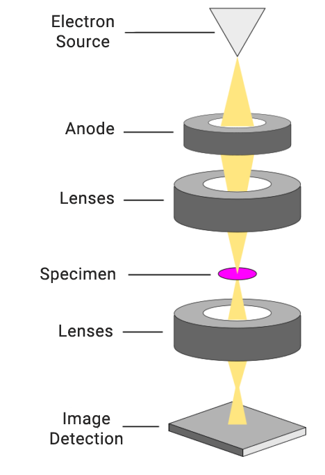
Image Detection :
Modern TEMs use advanced detectors, such as CCD cameras or other electronic sensors, to capture the magnified electron image.
Visualization and Analysis :
The captured image can be visualized on a screen for analysis and interpretation. Special techniques, like electron diffraction or energy-dispersive X-ray spectroscopy (EDS), can provide additional information about the specimen's composition and crystal structure.
Resolution and Magnification :
The resolution of a TEM is determined by the wavelength of electrons, allowing for extremely high magnifications and resolving power.
Biology and Medicine :
Cell Organelles Visualization :
TEM allows researchers to visualize and study various cell organelles, such as the nucleus, mitochondria, endoplasmic reticulum, Golgi apparatus, lysosomes, and peroxisomes, at a resolution sufficient to discern their internal structures and functions. This provides insights into cellular processes and organization.

Membrane Structures :
TEM is instrumental in studying the structure of cellular membranes, including the plasma membrane and various intracellular membranes. It helps researchers understand the composition, arrangement, and dynamics of lipid bilayers and associated proteins.
Cytoskeleton Examination :
The cytoskeleton, comprising microtubules, microfilaments, and intermediate filaments, plays a crucial role in cell structure and function. TEM enables detailed visualization of cytoskeletal elements, helping researchers understand their organization and involvement in processes like cell division and motility.
Virus and Pathogen Characterization :
TEM is widely used in virology and microbiology to study the ultrastructure of viruses, bacteria, and other pathogens. It provides detailed information about the morphology and replication strategies of these microorganisms, aiding in the development of antiviral and antibacterial strategies. TEMs assist pathologists in diagnosing diseases by providing high-resolution images of tissues and cellular abnormalities.

Cellular Junctions and Interactions :
TEM allows for the examination of cellular junctions, such as tight junctions, desmosomes, and gap junctions. Understanding these structures is crucial for insights into cell-cell interactions, tissue architecture, and the maintenance of epithelial integrity.
Subcellular Compartmentalization :
TEM is essential for investigating how cells compartmentalize various biochemical processes. For instance, it helps in understanding how enzymes and substrates are localized within specific organelles and how these compartments contribute to cellular function.
Neuronal Ultrastructure :
In neuroscience, TEM is employed to study the ultrastructure of neurons, synapses, and associated structures. This helps in understanding synaptic transmission, neural connectivity, and the morphological basis of neurological diseases.
Materials Science :
Crystallography and Microstructure Analysis :
TEM allows for the visualization of crystal structures at an atomic or near-atomic resolution. This is crucial for determining the microstructure of materials, including grain boundaries, defects, and dislocations. Researchers use TEM to study the arrangement of atoms in various materials, providing insights into their mechanical and electronic properties.
Nanoparticle Characterization :
TEM is employed to characterize nanoparticles and nanomaterials. It helps researchers determine the size, shape, and distribution of nanoparticles, which is essential for understanding their properties and optimizing their use in various applications, such as catalysis and drug delivery.
Defect Analysis :
TEM is a powerful tool for studying defects in materials, including vacancies, dislocations, and stacking faults. Examining these defects at the atomic level provides information about material strength, conductivity, and other mechanical properties.
Phase Identification :
TEM is used to identify different phases within a material. This is particularly important in multi-phase materials or composite materials where understanding the distribution and interactions of different phases is critical for optimizing performance.
Material Deformation and Fracture Studies :
TEM is employed to investigate the mechanisms of material deformation and fracture at the atomic and nanoscale. This information is crucial for understanding the behavior of materials under stress and for designing materials with enhanced mechanical properties.
Polymer and Composite Characterization :
In the study of polymers and composites, TEM provides detailed information about the morphology, dispersion of components, and interfacial interactions. This is valuable for optimizing the properties of materials used in industries ranging from aerospace to electronics.
Catalyst Characterization :
TEM is widely used in catalysis research to study the structure of catalyst nanoparticles. Understanding the morphology and surface properties of catalysts at the nanoscale is essential for improving catalytic efficiency and selectivity in chemical processes.
Electronic and Magnetic Properties :
TEM, especially high-resolution TEM (HRTEM), is employed to investigate the electronic and magnetic properties of materials. It can provide insights into the arrangement of atoms and electron distribution, aiding the development of materials for electronic and magnetic applications.
Quantum Dot and Nanowire Studies :
TEM is instrumental in the study of nanoscale materials like quantum dots and nanowires. Researchers use TEM to characterize the size, shape, and crystal structure of these materials, which are important for applications in electronics and photonics.
Physics :
Quantum Dots and Nanowires :
Researchers use TEM to investigate the properties of quantum dots and nanowires, which are essential components in the development of nanoelectronic devices and quantum technologies.

Condensed Matter Physics :
TEM is crucial in condensed matter physics for studying the behavior of materials in various states, such as liquids, solids, and gases. It provides insights into phenomena like superconductivity, magnetism, and electronic properties.
Nanotechnology :
In the field of nanotechnology, TEM plays a central role in characterizing nanomaterials, including nanoparticles, nanowires, and nanotubes. It aids in understanding the size, shape, and arrangement of atoms in these structures.
Low-Dimensional Systems :
TEM is employed to study low-dimensional systems like graphene and other two-dimensional materials. It helps physicists explore the unique electronic and mechanical properties of these materials.
Electron Diffraction Studies :
TEM can be used for electron diffraction studies, enabling researchers to analyze the crystal structure of materials. This information is crucial for understanding the symmetry and arrangement of atoms in crystals.
Plasmonics and Photonics :
In the field of plasmonics and photonics, TEM helps physicists investigate the interaction of light with metallic nanoparticles and nanostructures. It is valuable for designing devices with enhanced optical properties.
Magnetic Materials :
TEM is employed to study magnetic materials at the atomic level. It aids in understanding magnetic structures, domain configurations, and the interactions between magnetic nanoparticles.
High-Pressure Studies :
TEM can be applied in high-pressure studies, allowing physicists to investigate the behavior of materials under extreme pressure conditions. This is important for understanding the properties of materials in planetary interiors or during compression.
Geology and Earth Sciences :
Mineralogy and Petrology :
TEM is used to study the crystal structure, defects, and composition of minerals. It aids in identifying different mineral phases, understanding mineralogical transformations, and characterizing mineral-hosted inclusions.

Clay Mineral Analysis :
TEM is instrumental in the analysis of clay minerals, providing detailed information about their crystal structure, layer stacking, and interlayer composition. This is crucial for understanding soil properties, sedimentary processes, and the behavior of clays in geological formations.
Characterization of Nanoparticles in Soils:
TEM allows for the characterization of nanoparticles in soils, including colloidal particles and nanoscale minerals. This is important for studying soil structure, nutrient cycling, and the mobility of contaminants.
Studies of Sedimentary Rocks :
TEM is employed to investigate the microstructure of sedimentary rocks, helping researchers understand diagenetic processes, cementation, and the distribution of minerals within the rock matrix.
Analysis of Volcanic Materials :
TEM is used to study volcanic rocks and ash, providing insights into the crystallinity, composition, and textures of volcanic minerals. This aids in understanding volcanic processes, eruption dynamics, and magma evolution.
Characterization of Ore Minerals :
TEM is applied in the study of ore minerals to determine their microstructure, mineral associations, and textures. This is important for ore exploration and understanding the genesis of mineral deposits.
Identification of Microfossils :
TEM is used in the identification and characterization of microfossils, providing insights into the morphology and composition of ancient life forms. This is important for paleontological and stratigraphic studies.
Analysis of Sediment Particles :
TEM is utilized to study the fine-grained particles in sediments, helping researchers understand sedimentary processes, transport mechanisms, and the provenance of sedimentary materials.
Environmental Science :
Particulate Matter Analysis :
TEM is used to analyze particulate matter in air and water samples. It helps identify and characterize nanoparticles, allowing researchers to understand the composition and morphology of particulates, which is critical for assessing air and water quality.
Soil Structure and Composition :
TEM aids in the study of soil structure and mineral composition at the nanoscale. It helps researchers understand soil processes, nutrient availability, and the interactions between soil components and contaminants.

Analysis of Environmental Contaminants :
TEM is employed to investigate the fate and transport of environmental contaminants at the nanoscale. This includes the study of contaminant interactions with soil particles, sediments, and aquatic systems.
Microplastics Characterization :
TEM is used to characterize microplastics in environmental samples. It provides information on the size, shape, and surface properties of microplastic particles, aiding in understanding their impact on ecosystems.
Study of Atmospheric Aerosols :
TEM helps analyze the composition and morphology of atmospheric aerosols, including natural and anthropogenic particles. This information is crucial for assessing the role of aerosols in climate change, air quality, and human health.
Water Quality Assessment :
TEM contributes to water quality assessment by providing detailed information on the structure and composition of suspended particles in water. This is important for understanding the impact of human activities on aquatic ecosystems.
Analysis of Environmental Microorganisms :
EM aids in the study of environmental microorganisms at the nanoscale. It provides information on microbial cell structures, interactions with minerals, and the formation of microbial biofilms in natural environments.
Carbon Nanomaterials in the Environment:
TEM is employed to study the distribution and behavior of carbon nanomaterials, such as carbon nanotubes and graphene, in environmental matrices. This is important for assessing the potential environmental impact of emerging nanotechnologies.


| Resolution (nm) at 80 kV | |
|---|---|
| Points | 0.50 nm |
| Lattice | 0.34 nm |
| Acceleration Voltage | |
| Range | 50kV, 80kV |
| Stability | 8×10⁻⁶ |
| Magnification | |
| Range | 1X to 400,000X |
| Steps | 16 |
| Diffraction | |
| Selected area | >1.5µm diameter, selected apertures (SAD) |
| >3 µm diameter, selected by micro-beam illumination | |
| Illumination system | |
| Electron Gun | Pre-centered tungsten hairpin filament |
| Beam Alignment | Electromagnetic beam alignment system |
| Double condenser | Factory aligned double condenser system specimen |
| Illumination adjustable from 3µm to 2mm diameter | |
| Objective lens | |
| Focal length | 2.6mm |
| Spherical aberration | 2.2mm |
| Chromatic aberration | 1.7mm |
| Astigmatism | Greater than 1µm |
| Projector lenses | |
| Number of lenses | 3 factory aligned electromagnetic projector lenses |
| Stability | 6×10⁻⁶ |
| Imaging modes | |
| Darkfield | Available |
| Bright-field | Available |
| SAD | Available |
| Vacuum system | |
| Vacuum pumps | Rotary and turbo molecular |
| Vacuum gauge | Pirani and cold cathode |
| Vacuum pressure | High vacuum pressure down to 10-6 mbar |
| Movements | |
| X-Y Sample Movements | Motorized |
| Sample tilt | Optional (details can be customized) |
| Apertures | Motorized |
| Software | |
| Imaging | Available |
| Image processing | Available |
| Apertures control | Available |
| X-Y Sample control | Available |
| Vacuum control | Available |
| Digital imaging system | |
| Motorized control of the scintillator | |
| Optical zoom of 10 X | |
| 16.0MP high resolution sensor (4608H × 3456V) | |
| Up to 25 frames per second | |
| High-speed data transmission | |