Paper summary
This article reviews atomic force microscopy (AFM) studies of DNA structure and dynamics and protein–DNA complexes, including recent advances in the visualization of protein–DNA complexes.

English
This article reviews atomic force microscopy (AFM) studies of DNA structure and dynamics and protein–DNA complexes, including recent advances in the visualization of protein–DNA complexes.
As a topographical technique, Atomic Force Microscopy (AFM) needs to establish direct interactions between a given sample and the measurement probe in order to create imaging information. The elucidation of internal features of organisms, tissues and cells by AFM has therefore been a challenging process in the past. The study at hand shows the applicability of the proposed protocol to exemplary biological samples, the resolution currently allowed by the approach as well as advantages and shortcomings compared to classical ultrastructural microscopic techniques like electron microscopy.
DNA carries the genetic information inside the cells and represents a sensitive target of ionizing radiation. Ionizing radiations induce free radicals in DNA constituents and thereby produce various types of DNA lesions such as base damage, DNA single-strand breaks (SSBs), DNA doublestrand breaks (DSBs), and DNA-protein crosslinks Ionizing radiation produces clustered DNA damage that contains two or more lesions in 10–20 bp. It is believed that the complexity of clustered damage (i.e., the number of lesions per damage site) is related to the biological severity of ionizing radiation. However, only simple clustered damage containing two vicinal lesions has been demonstrated experimentally. Here we developed a novel method to analyze the complexity of clustered DNA damage by AFM.
Atomic force microscopy (AFM)-based methods have matured into a powerful nanoscopic platform, enabling the characterization of a wide range of biological and synthetic biointerfaces ranging from tissues, cells, membranes, proteins, nucleic acids and functional materials. AFM-based force spectroscopy allows their mechanical, chemical, conductive or electrostatic, and biological properties to be probed. The combination of AFM-based imaging and spectroscopy structurally maps these properties and allows their 3D manipulation with molecular precision. In this Review, we survey basic and advanced AFM-related approaches and evaluate their unique advantages and limitations in imaging, sensing, parameterizing and designing biointerfaces.
Electrochemical AFM is a powerful tool for the real-space characterization of catalysts under realistic electrochemical CO2 reduction (CO2RR) conditions. The evolution of structural features ranging from the micrometer to the atomic scale could be resolved during CO2RR. Using Cu(100) as model surface, distinct nanoscale surface morphologies and their potential-dependent transformations from granular to smoothly curved mound-pit surfaces or structures with rectangular terraces are revealed during CO2RR in 0.1 m KHCO3. The density of undercoordinated copper sites during CO2RR is shown to increase with decreasing potential. In situ atomic-scale imaging reveals specific adsorption occurring at distinct cathodic potentials impacting the observed catalyst structure. These results show the complex interrelation of the morphology, structure, defect density, applied potential, and electrolyte in copper CO2RR catalysts.
Weakly bound, physisorbed hydrocarbons could in principle provide a similar waterrepellency as obtained by chemisorption of strongly bound hydrophobic molecules at surfaces.
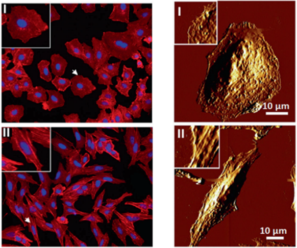
Mechanics are intrinsic properties which appears throughout the formation, development, and aging processes of biological systems. Mechanics have been shown to play important roles in regulating the development and metastasis of tumors, and understanding tumor mechanics has emerged as a promising way to reveal the underlying mechanisms guiding tumor behaviors. AFM has appeared as an excellent platform enabling simultaneously characterizing the structures and mechanical properties of living biological systems ranging from individual molecules and cells to tissue samples with unprecedented spatiotemporal resolution, offering novel possibilities for understanding tumor physics and contributing much to the studies of cancer.

Weakly bound, physisorbed hydrocarbons could in principle provide a similar waterrepellency as obtained by chemisorption of strongly bound hydrophobic molecules at surfaces.
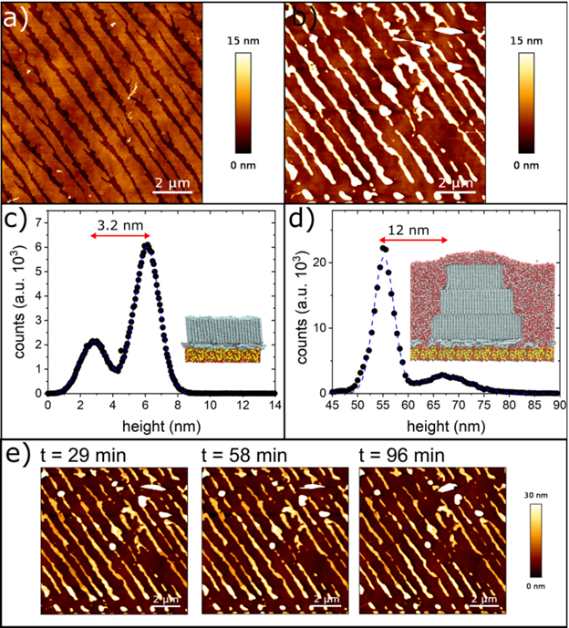
Experiments: Here we present experiments and computer simulations on the wetting behaviour of water on molecularly thin, self-assembled alkane carpets of dotriacontane (n-C32H66 or C32) physisorbed on the hydrophilic native oxide layer of silicon surfaces during dip-coating from a binary alkane solution.
By changing the dip-coating velocity we control the initial C32 surface coverage and achieve distinct film morphologies, encompassing homogeneous coatings with self-organised nanopatterns that range from dendritic nano-islands to stripes. We found out that both the coverage and morphology of the nanopatterns can be controlled by the sample preparation velocity.
we find a coating transition at 0.83 mm/s between two types of organic growth structure, which is reflected in both the surface coverage as well as the fractal dimension of the resulting nanopatterns. The transition between the two coating regimes affects the wetting properties of water, the measured contact angles also group into two regimes.

Integrated photoelectrochemical devices rely on the synergy between components to efficiently generate sustainable fuels from sunlight. The micro- and/or nanoscale characteristics of the components and their interfaces often control critical processes of the device, such as charge-carrier generation, electron and ion transport, surface potentials, and electrocatalysis. in this paper we are going to study spatial properties and structure–property relationships of these which can provide insight into designing scalable and efficient solar fuel components and systems.

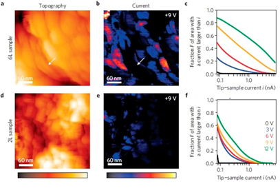
Intermolecular single-electron transfer on electrically insulating films is a key process in molecular electronics and an important example of a redox reaction. Electron-transfer rates in molecular systems depend on a few fundamental parameters, such as interadsorbate distance, temperature, and, in particular, the Marcus reorganization energy. Here, we investigate redox reactions of single phthalocyanine (NPC) molecules on multilayered NaCl films. Employing the atomic force microscope as an ultralow current meter allows us to measure the differential conductance related to transitions between two charge states in both directions. Our approach provides insight into single-electron intermolecular transport.
This article is about surface analysis of pure aluminum thin films by AFM and films were deposited on stainless and mild steel substrates through rf magnetron sputtering at rf powers of 150 and 200 W and the result of this article shows that the morphologies of the surface structures of Al films vary with power and substrate type.